DNA Microarrays
October 26, 2021 | 4 min read

October 26, 2021 | 4 min read

There are about 200 different cell types in the human body alone, all with the same genetic material in the form of DNA. Early evidence showed that from about 25,000 genes seen in humans, only a fraction are expressed at a given time, in a given cell. Naturally, this differential gene expression across various cells in an organism has fascinated scientists for years. DNA microarrays was a technique that was developed in the mid-1990s to measure the expression levels of several genes simultaneously in a single reaction efficiently. Understanding the expression of genes is integral to fully comprehend the fundamental aspects underlying several physiological processes.
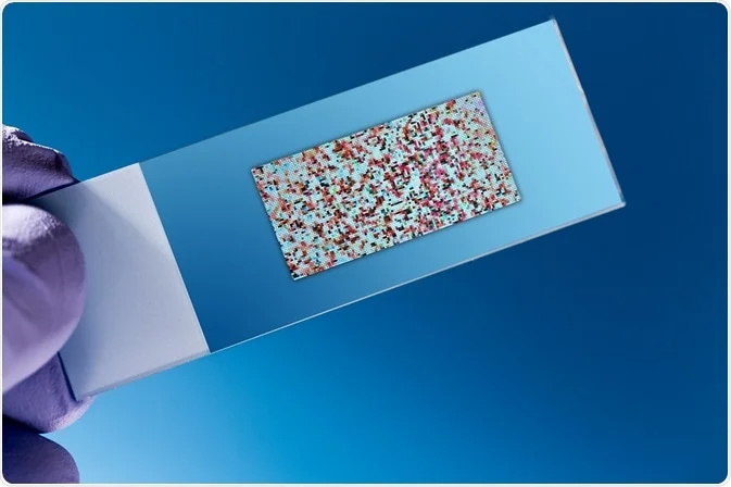
A large number of spotted samples, known as probes, whose identities are clearly known are immobilized on a solid support surface. The solid surface can be a glass, plastic or silicon biochip, otherwise known as a DNA chip or gene array. The probes can be DNA, cDNA, or oligonucleotides. This orderly arrangement of samples forms an array, where the sample spot sizes are typically less than 200 microns in diameter, and usually, thousands of sample spots are analyzed simultaneously.
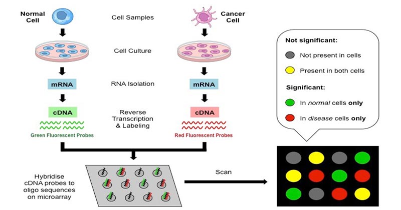
A typical protocol begins by obtaining complementary single-stranded DNA molecules. This can be done by extracting the messenger RNA (mRNA) from the cell of interest. The mRNA transcribed from the genetic sequence contains the information which is required for the synthesis of proteins by the ribosome.
The mRNA is reverse transcribed into complementary DNA (cDNA), which has sequences identical to the segments of the genes which produced the mRNA. During this process, the cDNA are usually labelled by incorporating a fluorescent dye in the DNA nucleotide, producing a fluorescent cDNA strand. If the starting material is limited, an additional amplification step can be used. The labelled cDNA are now added to the microarrays wherein it would get hybridized to its synthetic known DNA probes attached on the microarray. This is followed by a series of washes in order to remove any unbound cDNA molecules.
Now, the microarray plate is scanned by exciting the fluorescent tags present on the cDNA molecules by a laser such that the sequences bound to the probe on the plate will give out a distinct signal. The total strength of the signal depends on the amount of target sample bound to the probes. The detected signals are quantified and an image of the microarray is generated. From this, we can elucidate the expression level of various genes that are expressed in the sample.
Since its discovery, DNA microarray has been an indispensable part of functional genomics, since it allows for a better understanding of the differential gene expression in different cell types. Using this information, scientists have been able to create gene expression profiles for various cell types, in different organisms under varying conditions, including the normal versus diseased state. A single microarray experiment can generate a lot of information about thousands of genes. Microarrays have revolutionized our understanding of gene expression and provided a lot of insights to understand growth, development, disease prognosis and aided the discovery of novel genes and drug targets.

Figure 1. Source