Non-Invasive study Of Soft Tissue

March 3, 2021 | 3 min read

cover-image

  • Computer Tomography Scan (CT Scan).
    In CT scan, differences in the absorption of X-ray by the brain tissue are used to construct a three dimensional image (3D image). Here the dose of X-ray needed is very low.
  • picture

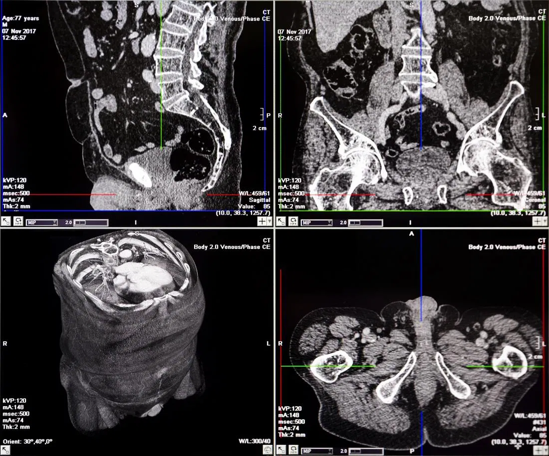
    Figure 1.A typical CT scan used to detect abnormalities in the soft tissue.


  • Magnetic Resonance Imaging (MRI).
    The principle of MRI is to use a magnetic field which shows differences in vibration of Protons of the water molecules present in the tissues of the body. This vibration of water molecules depends on the chemical surroundings of the tissue. Through MRI studies, many metabolic processes of the brain and other organs may be investigated. MRIs are used to detect the location of tumours, sites of hemorrhage, intracranial bleeding etc.
  • picture


  • Positron Emission Tomography (PET).
    PET scan is based on the use of positron, which is like an electron except that it has positive (+) charge. It also gives 3D images of where the molecules are located. It helps to measure the blood flow in the brain, glucose utilisation and oxygen consumption, and for the diagnosis of psychiatric disorders, brain tumours, epilepsy and degenerative changes due to Alzheimer’s disease.
  • picture

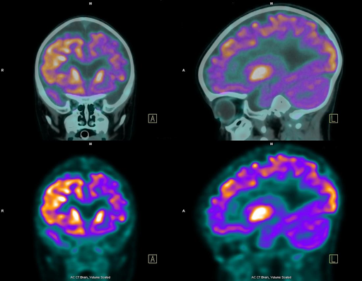
    Figure 3.PET images show the active cells as bright or ‘hot’ spots.


  • Ultrasound.
    A form of acoustic energy, or sound, that has a frequency that is higher than the level of human hearing.Ultrasound imaging uses sound waves to produce pictures of the inside of the body. Ultrasound waves are produced by a transducer, which can both emit ultrasound waves, as well as detect the ultrasound echoes reflected back. When these echoes hit the transducer, they generate electrical signals that are sent to the ultrasound scanner. Using the speed of sound and the time of each echo’s return, the scanner calculates the distance from the transducer to the tissue boundary. These distances are then used to generate two-dimensional images of tissues and organs.
  • picture

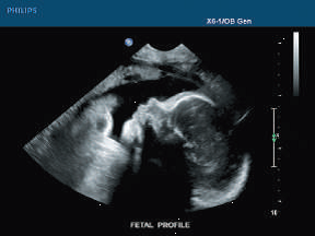
    Figure 4.Cross-section ultrasound image of a fetus Source


About This Author

Siddharth Shivanandan is a Batch 17 BS-MS student at IISER TVM