The Synchrotron
Oct 20, 2020 | 1 min read

Oct 20, 2020 | 1 min read

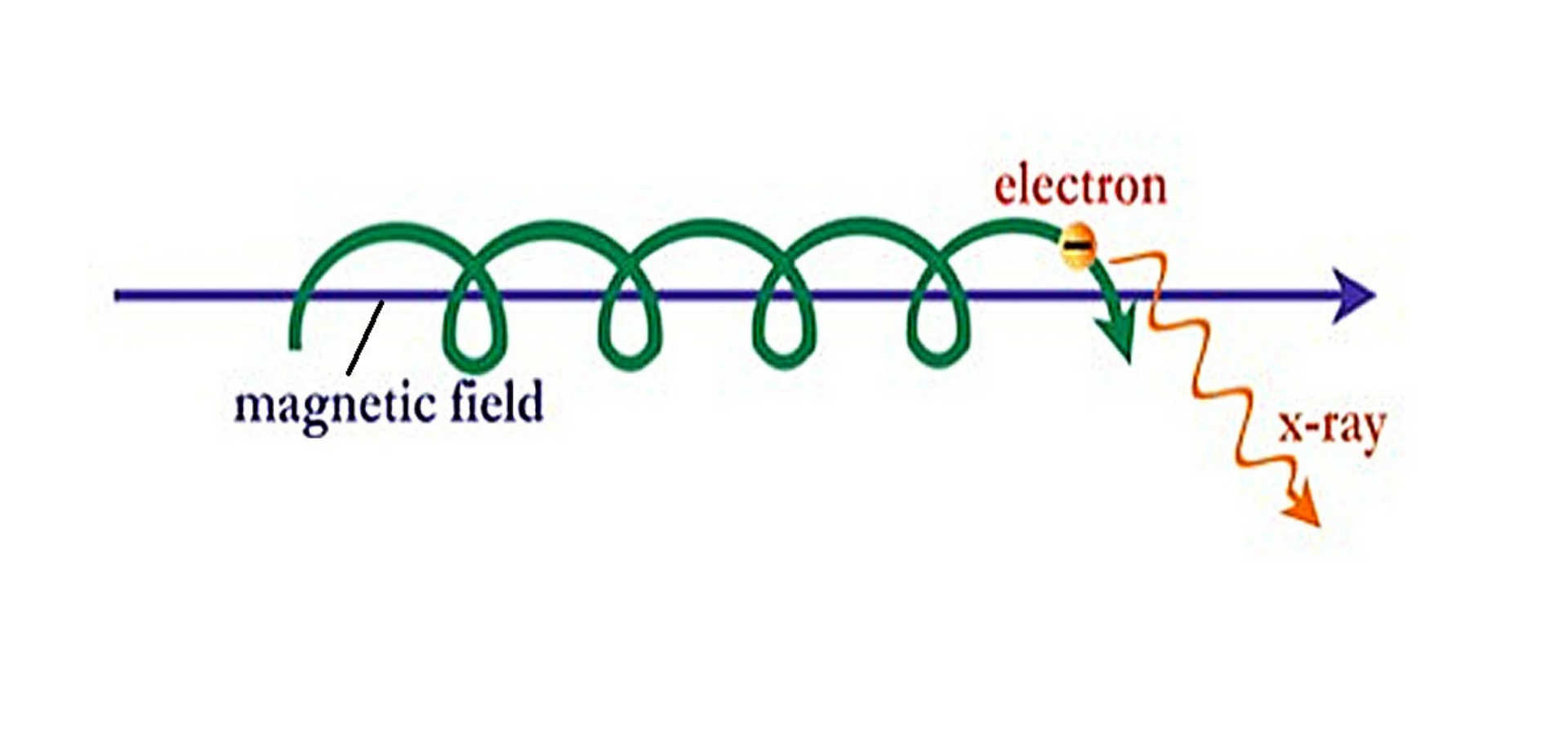
We have learnt this fact consistently. A particle that changes direction is an accelerating particle. A synchrotron takes advantage of this very basic concept. In a synchrotron, which is about the size of a football field, an electron charge
changes direction constantly and releases energy. When it begins to move fast enough the emitted energy is at X-ray wavelength.
A synchrotron thus exists to accelerate electrons to extremely high energy and then make them change direction periodically. The resulting X-rays are emitted as dozens of thin beams, each directed toward a beamline next to the accelerator.
The role of synchrotron radiation in structural biology has been growing rapidly over the last twenty years. A recent survey of the literature shows that synchrotron radiation was used in nearly half of the new structure determinations. The
unique properties of synchrotron light mean that experimental results are far superior in accuracy, clarity, specificity and timeliness to those obtained using conventional laboratory equipment. The broad range of available wavelengths allow
scientists to look at the size and shape of macromolecules and voids in bulk materials, peer into the biochemistry of single cells and delve all the way down to the bonds between atoms.
Originally, synchrotrons were used exclusively in the field of particle physics, allowing scientists to study collisions between subatomic particles with higher and higher energies. First-generation synchrotron light sources were basically
beamlines built onto existing facilities designed for particle physics studies. Second generation synchrotron light sources were dedicated to the production of synchrotron radiation and employed electron storage rings to harness the
synchrotron light.
Current (third generation) synchrotron light sources optimize the intensity of the light by incorporating long straight sections into the storage ring for ‘insertion devices’. Laboratories around the world are now working to overcome the
technical challenges associated with the development of fourth generation light sources, which are likely to utilise hard X-ray free-electron lasers (FEL).
An example of a synchrotron is The DIAMOND Light Source. This is the United Kingdoms national synchrtron. It works like a giant microscope, by producing bright light that scientists can use to study a vast range of speciments from fossils to
viruses and vaccines. DIAMOND stands for DIpole And Multipole Output for the Nation.

Figure 1. Synchrotron Radiation schematic. This shows us the X rays produced by an electron made to accelerate in a magnetic field. The magnetic field results in the electron moving in a spiral path. Hence, constantly accelerating.Source

Figure 2. DIAMOND Light Source, UK’s national synchrotron. DIAMOND stands for DIpole And Multipole Output for the Nation.