Arachnophobia Cut Out of Man's Brain
Oct 31, 2020 | 1 min read

Oct 31, 2020 | 1 min read
The amygdala, two almond shaped clusters of nuclei located right above the hippocampus in the frontal temporal lobe, are responsible for one’s ability to feel certain emotions, especially fear.
In 2014, a 44 year old businessman started having seizures out of the blue, and brain scans showed he had an abnormality in his left amygdala. Further studies showed that he had sarcoidosis which is a rare condition starting from the lungs
and eventually moving up to the brain. Medical experts decided it was time to remove the left amygdala. After the successful surgery, the same man who would once try to smash spiders with tennis balls was now fascinated by these tiny
harmless
creatures. While the man, who was also terrified of speaking in public, continued to feel the same way about it, somehow only the fear of spiders had been erased from his brain.
Nick Medford, at the Brighton and Sussex Medical School, UK suggested that the neural pathways related to panic-type fear response were eliminated with the removal of the left amygdala,while parts of the amygdala responsible for generalised
fear remained intact. Other aspects of the man’s panic response could not be assessed as he had no other phobias and refused to undergo further testing.
The amygdala is too deep in the brain to attempt to disrupt it using non-invasive techniques to cure other people of their phobias, says Medford. But there are several other fear-dampening techniques being trialled, including weakening
phobias using a blood pressure pill and stimulating certain brain areas in a bid to erase fearful memories.
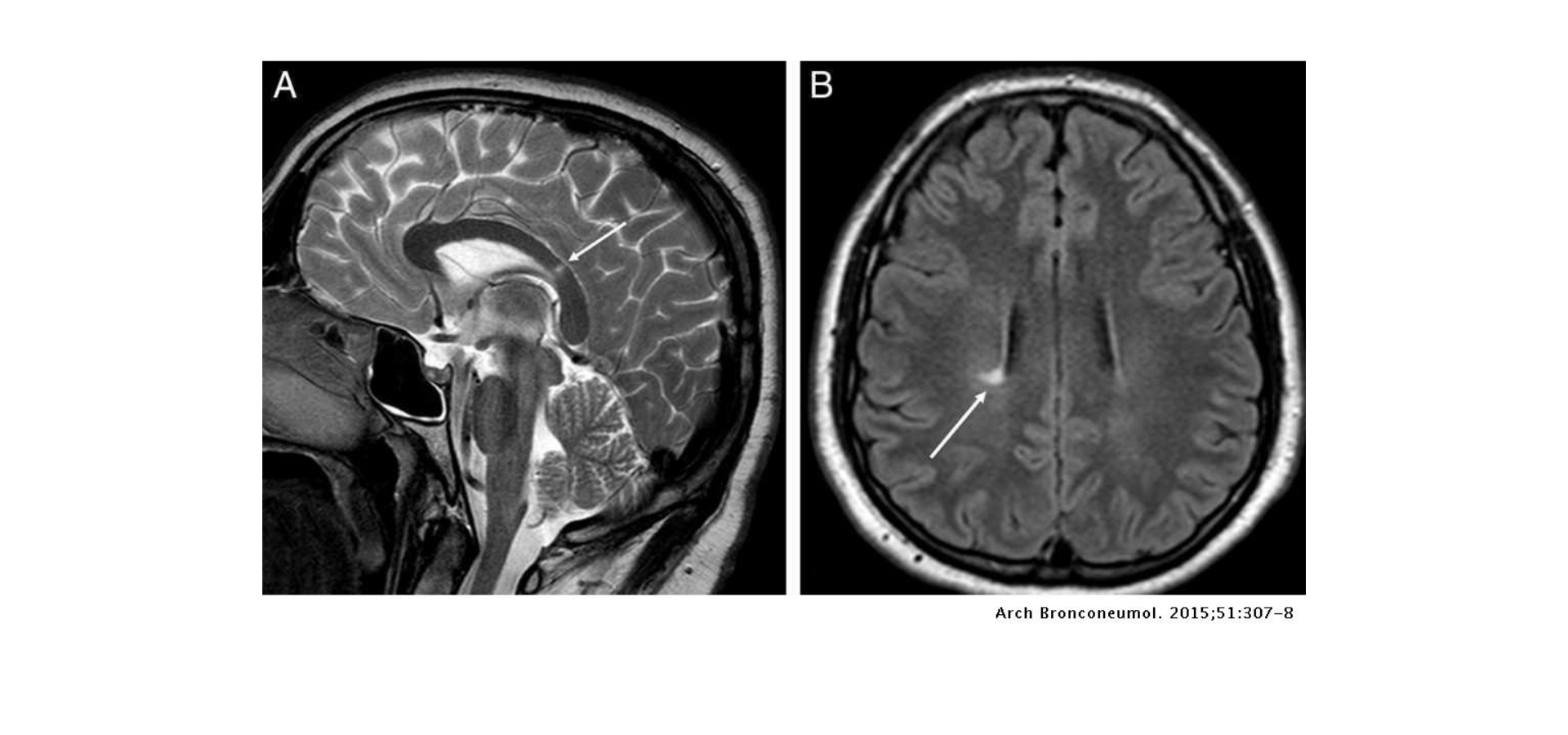
Figure 1. Magnetic resonance imaging (MRI) of the head in T2 (A) showing a hyperintense focal lesion in the splenium of the corpus callosum (white arrow). Axial FLAIR image (B) showing a hyperintense focal lesion in the periventricular white matter (white arrow). Both lesions are radiologically non-specific, but in the right clinical context can be indicative of demyelinating disease. Source
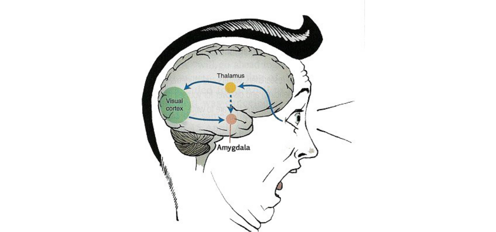
Figure 2. The amygdala is a small structure located within the medial temporal lobes (MTL), consisting of a discrete set of nuclei. It has a reputation as the “fear center” or “emotion center” of the brain. Source

Oct 16, 2020 | 2 min read

Nov 25, 2020 | 4 min read